Advanced Microscopy

We're here to support your research with our advanced microscopy expertise.
It is our mission to facilitate science and provide access to know-how and measurement time on state-of-the-art microscopes. We have ample devices available, including numerous confocal microscopes and super-resolution systems, slide scanners, high content screening, and 3D whole organ imaging (e.g. light sheet microscopes) as well as transmission and scanning electron microscopes, light and electron microscopy-combined systems and block face scanning electron microscopy solutions. We take pride in our support for image analysis both with infrastructure and expertise.
VIB Bioimaging Core
The VIB Bioimaging Core in Leuven supports imaging from in vitro to in vivo, from the meso to the Ångström scale, and from morphology to mechanism. They have ample solutions available, including confocal microscopes, slide scanning, high content screening, and 3D whole organ imaging.
Take a Virtual Tour
Want to see our microscopes? Check out what we have available at the VIB-KU Leuven Center for Brain & Disease Research via our 360° virtual tour.
In the Spotlight
A selection of research papers from the VIB-KU Leuven Center for Brain & Disease Research labs which utilised our advanced microscopy technologies.
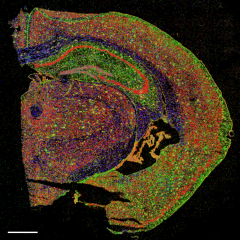
Spatial Transcriptomics and In Situ Sequencing to Study Alzheimer’s Disease
Published in Cell
From the Bart De Strooper Lab
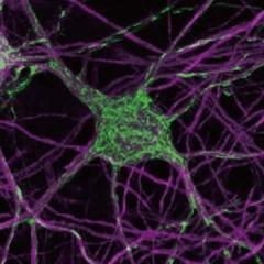
Mitochondria metabolism sets the species-specific tempo of neuronal development
Published in Science
From the Pierre Vanderhaeghen Lab
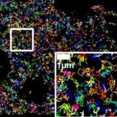
Safe targeting of T cell acute lymphoblastic leukemia by pathology-specific NOTCH inhibition
Published in eLife
From the Lucía Chávez-Gutiérrez Lab
Stay up-to-date

We are a Nikon Center of Excellence

In Vivo Advanced Imaging

Use Our Microscopy Facilities
